
Nanoscale imaging CRO · Preclinical R&D
Quantitative tissue imaging at subcellular level
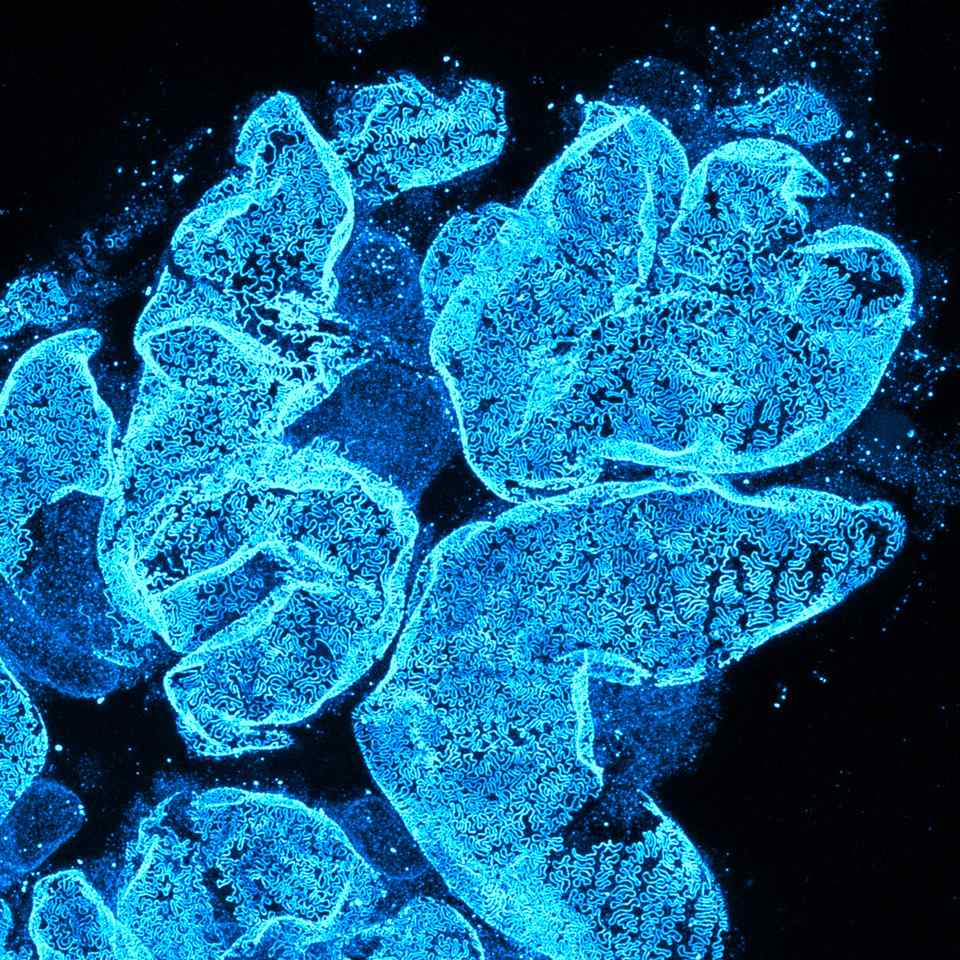
Magnephy resolves drug localisation and tissue response at the nanoscale, in 3D. Reproducible, AI-quantified data, ready for preclinical programs.
- 30 nm
- spatial resolution
- 3D
- volumetric readout
- AI
- automated quantification
- 10+ years
- advanced imaging experience




What we deliver
Drug-target colocalisation.
Drug, target, and structure resolved in the same volume, at 30 nm.
Learn more →Subcellular biodistribution.
Biomarker and spatial drug distribution, across whole tissue volumes, in 3D.
Learn more →Quantitative pathology.
Evaluate tissue response down to the subcellular level with AI-quantified data.
Learn more →One workflow.Three steps.One quantitative readout.
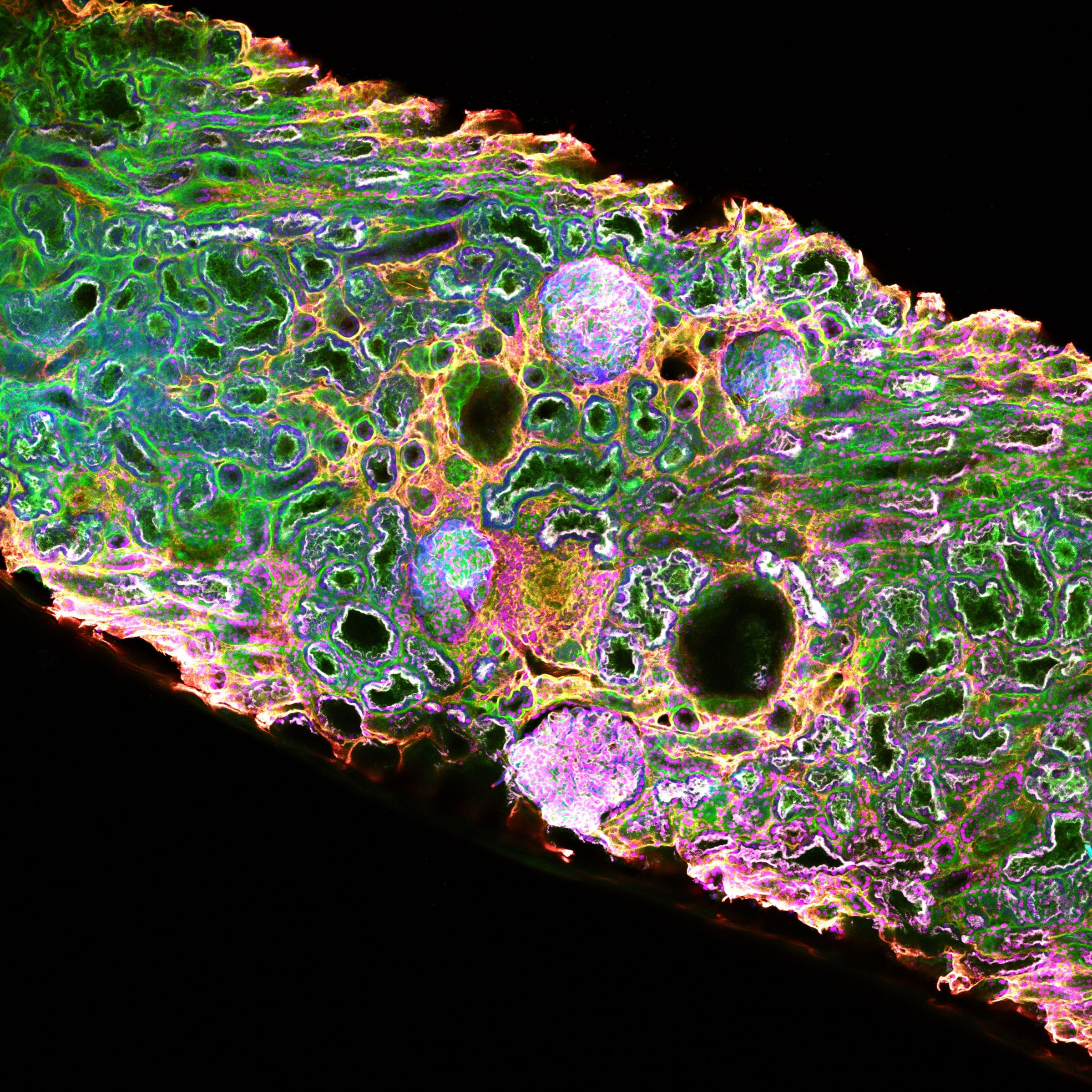
Tissue clearing
Intact biopsy, optically transparent.
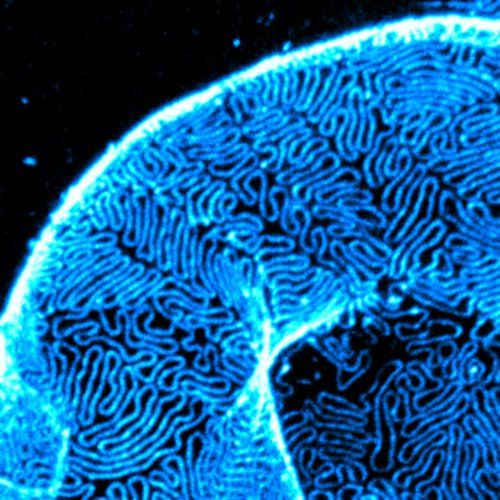
Super-resolution imaging
STED at the nanoscale, in 3D.
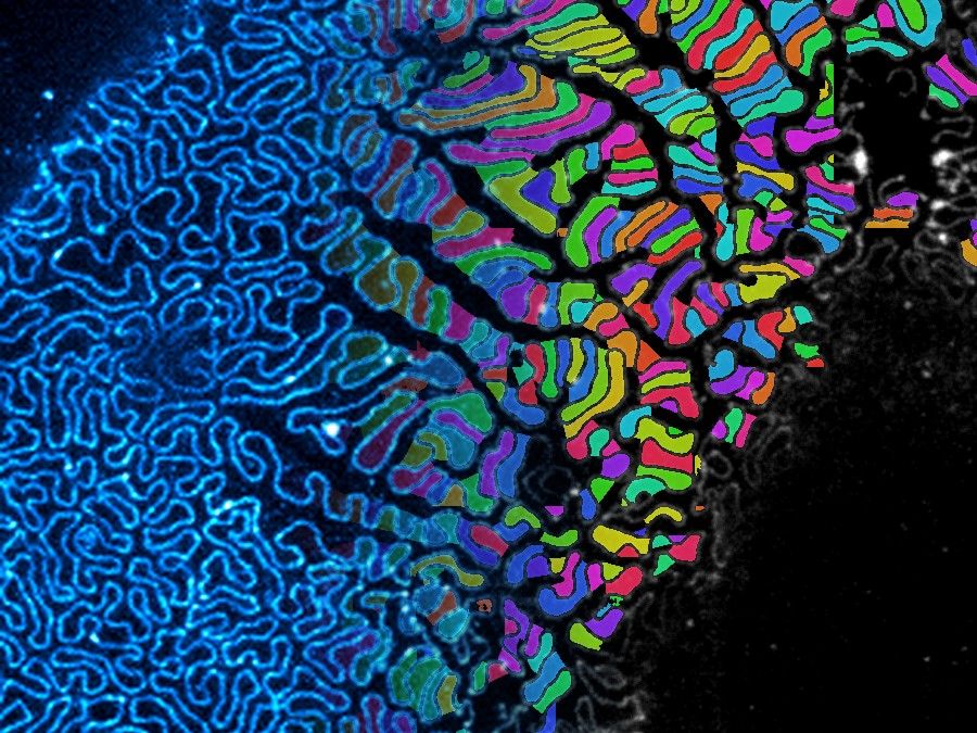
AI segmentation
Reproducible, quantitative readouts.
Achieving the best results with any imaging platform depends as much on optimal sample preparation as having the right microscope. At Magnephy we make great results, simple. Tissue clearing, STED super-resolution, and AI segmentation, integrated into a single validated workflow for perfect results, every time.
Read the method →Applications
Where the platform is used today.
Quantitative kidney pathology
Evaluate foot-process effacement, slit-diaphragm density, glomerular basement membrane and immune deposits. CKD, FSGS, IgA nephropathy, diabetic and lupus nephritis.
Read more →Antibody therapeutics
Drug-target colocalisation and biodistribution, in 3D, across tissues and organs.
Read more →In development
We also build specialised assays per program, extending the same quantitative pathology and biodistribution to other diseases and drug modalities.
Tell us what we should develop next →Proven science
Built on 10+ years of research at leading institutions.
The platform was developed, published, and validated over a decade of peer-reviewed research. From Stockholm, Magnephy brings this technology to industrial standards, delivering decision-grade data to preclinical and clinical-stage programs in the EU and US.
First to visualise the slit diaphragm by light microscopy.
Our co-founders were first to visualise the slit-diaphragm protein architecture in optically cleared kidney tissue, the breakthrough the platform is built on.
Proven in academia, adapted for industry.
Developed and published across a decade of peer-reviewed academic research, then optimized in-house for the throughput, reproducibility, and rigor that industrial programs demand.
Built on super-resolution and tissue clearing.
The platform combines optical tissue clearing, STED super-resolution, and AI quantification, refined over more than a decade of advanced imaging.
The results far exceeded our most optimistic expectations… The images Magnephy generated were breathtaking, providing a truly intuitive understanding of the underlying biology.

How a project works
From sample-in-lab to decision-grade data.
Plan
Our scientists help you plan the optimal assay for your question, model, and program stage.
Run
Validated assays return results in 2 to 4 weeks. Biomarker validation in 4 to 8 weeks. Specialised assays can be set up to milestones.
Results
Quantitative readouts, 3D visualisations and reporting tailored to your needs.