
Platform
Imaging at the scale of mechanism.
Magnephy resolves drug, target, and tissue structure at 30 nm in 3D, in intact tissue. Nanoscale 3D spatial biology, with reproducible AI-quantified readouts of targeting, biodistribution, and tissue response, ready for preclinical and translational decisions.
The bottleneck
The real bottleneck is the sample, not the microscope.
In tissue imaging, the resolution you achieve is set by the sample. Optical scattering, labelling efficiency, and sectioning artefacts cap the value of every downstream technique. Magnephy's tissue clearing and labelling protocols, refined over more than a decade, remove that cap, so biology can be measured with nanoscale precision in intact tissue.
Workflow
One workflow.Three steps.One quantitative readout.
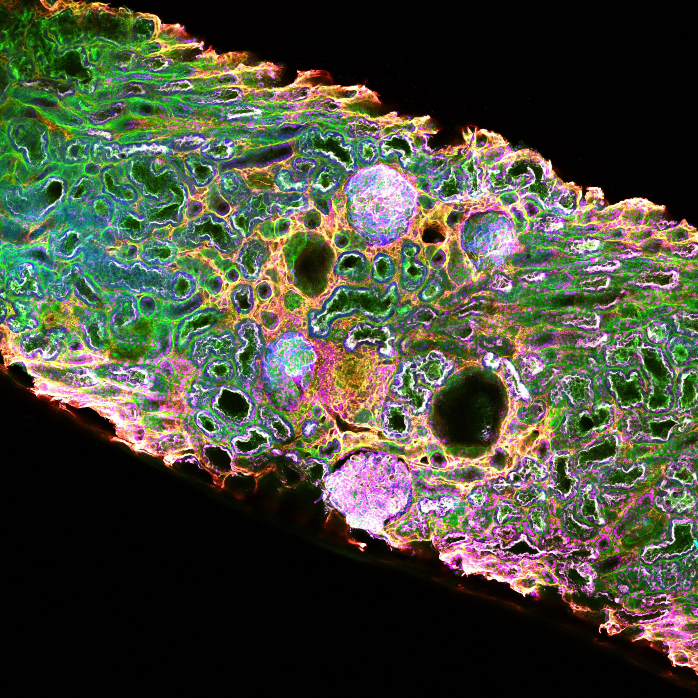
Tissue clearing
Optical clearing turns intact biopsies and whole-tissue samples transparent without sectioning. Refined over a decade at SciLifeLab and KTH Royal Institute of Technology; published in three Kidney International papers (2016, 2021, 2023).
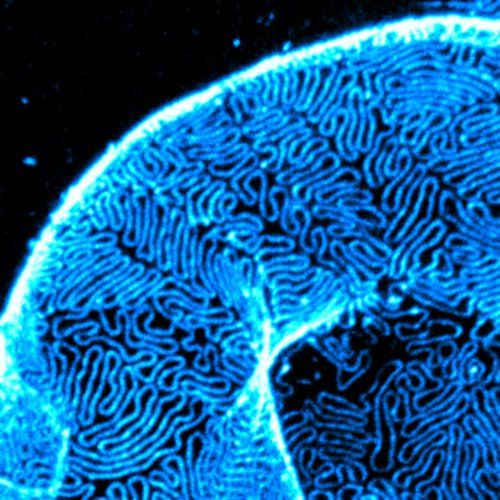
Super-resolution imaging
STED super-resolution microscopy resolves structure at 30 nm in 3D. The platform reads inside individual cells: organelle-level biodistribution, drug-target colocalisation and subcellular biomarker counts.
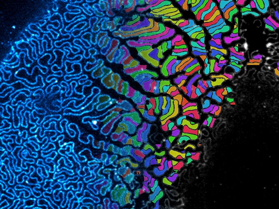
AI segmentation
AI-driven segmentation turns volumetric image stacks into reproducible quantitative readouts: per-cell metrics, per-region distributions and per-cohort comparisons.
Capabilities
What you actually get back.
Drug-target colocalisation
Where the therapeutic sits relative to its target, at 30 nm in 3D. Direct, not inferred. Quantified across whole 3D volumes.
Subcellular biodistribution
Where the therapeutic ends up inside individual cells. Tissue penetration and target-site exposure, resolved as organelle-level distribution in real tissue, not approximated by proximity.
Quantitative pathology
Tissue structure, disease state and treatment response, AI-quantified to replace qualitative scoring. Specialized assays of podocyte and glomerular pathology for nephrology programs are available.
Multiplex biomarker readouts
Multi-channel marker counts and spatial distributions across whole tissue volumes. Reproducible across samples and timepoints, comparable across cohorts.
Comparison
How the platform differs from standard tissue imaging.
Conventional methods trade resolution against molecular detail against 3D context. Magnephy combines tissue clearing, super-resolution imaging, and AI segmentation into a single workflow that achieves all three at once, in intact tissue.
| Confocal IF · EM · IHC | Magnephy |
|---|---|
| 2D sections | 3D whole tissue volumes |
| Resolution OR molecular detail OR throughput | All three in one workflow |
| Drug-target colocalisation inferred at >300 nm | Drug-target colocalisation resolved at 30 nm |
| Manually-scored, qualitative | AI-segmented, quantitative, reproducible |
| Each technique a separate run | Single integrated platform |